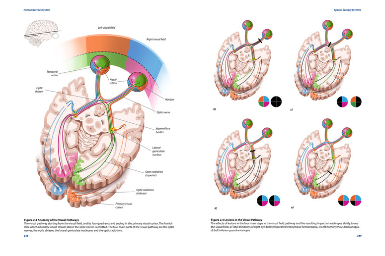
QCVISUAL
  • HOME
  • PORTFOLIO
  • eLearning
  • ABOUT
  • CONTACT

Anatomy of the Visual Pathway and Lesions

Picture
Back to Gallery
DESCription
Title

Medium

Audience

Format

Supervisors

Purpose
Anatomy and lesions of the visual pathways

Adobe Photoshop and Illustrator CS6

Medical and neuroanatomy students

Print

Prof. L. Wilson-Pauwels, TA Andrea Gauthier, Content expert: Dr. M. Wiley

This purpose of this textbook illustration is to explain the anatomy of the visual pathway from the retina to the primary visual cortex and the accompany effects on the visual field when lesions occur in the pathway. The technical goals of this project included rendering skills, layout, and following the workflow and requirements of textbook publications.


Process Work

My first sketches included some basic color to separate the visual fields and tracts. At this stage, I had many illustrations and wanted to approach this topic in multiple ways. However, there was too much visual information and they didn't work coherently with each other, so I pared it down and chose the best way to focus on conveying the information I wanted. 
Picture
Picture
I wanted to approach the visual pathways using a brain slice for both the pathway and lesions, and tried to work on a layout that fit into one page. As I progressed and rendered my sketch out more, I realized that the images were too small. Therefore, for my final version, I arranged it into a two-page textbook layout using the specifications given. 
Picture

References

AFIP #406908 Pilocytic astrocytoma (Optic glioma). http://commons.wikimedia.org/wiki/Pilocytic_astrocytoma
England, M. A., & Wakely, J. (2006).Color atlas of the brain and spinal cord: an introduction to normal neuroanatomy(2nd ed.). Philadelphia, PA: Mosby Elsevier.
Kandel, E. R., Schwartz, J. H., & Jessell, T. M. (2000). Principles of neural science(4th ed.). New York: McGraw-Hill, Health Professions Division.
Kiernan, J. A., & Barr, M. L. (20082009).Barr's the human nervous system: an anatomical viewpoint (9th ed.). Philadelphia: Lippincott, Williams & Wilkins.
Martini, F., Timmons, M. J., & Tallitsch, R. B. (2012). Human anatomy (6th ed.). Boston: Pearson Benjamin Cummings.
Nolte, J (2009). The Human Brain an Introduction to its Functional Anatomy.  Philadelphia PA: Elsevier.
Pauwels, L., & Akesson, E. J. (2002).Cranial nerves (2nd ed.). Hamilton, Ont.: B C Decker.
Poritsky, R. (1984). Neuroanatomical pathways. Philadelphia: Saunders.
Purves D, Augstine G A, Fitzpatrick D, Hall W, LaMantia A-S, McNamara J O, Williams S M (2007). Neuroscience 4th Ed. Sunderland: MA.Sinauer Associates.
Rubin M, Safdieh J (2007). Netter’s Concise Neuroanatomy. Philadelphia PA: Elsevier
Wilson-Pauwels L, Akesson E J, Stewart P A, Spacey D (2002). Cranial Nerves in Health and Disease.  BC: Decker Inc.
Wilson-Pauwels L, Akesson E J, Stewart P A, Spacey D (2010). Cranial Nerves 3rd Ed.  PMPH-USA.

Services

Medical Illustration
Graphic Design
Animation
Scientific Illustration

Pages

Home
Portfolio
About

Contact
QCVisual medical illustration logo
  • HOME
  • PORTFOLIO
  • eLearning
  • ABOUT
  • CONTACT